Human brain cells grafted into mouse cortex is science fiction no more
Stanford researchers have grafted human brain organoids into the cortices of living mice — a first that opens new doors for disease modeling and raises urgent ethical questions the scientific world has been avoiding.
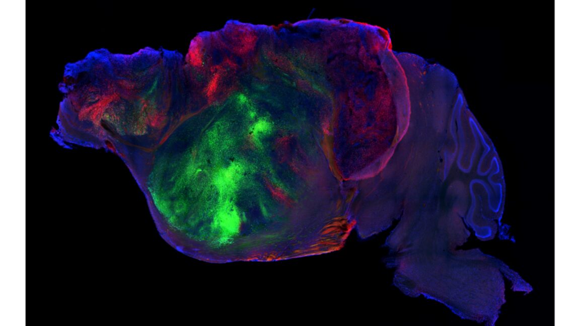
A green line in the gray matter
Under a fluorescence microscope, the image is striking in its simplicity: green processes — extensions of human-derived neurons — reaching into the tissue of a mouse brain. This is not an abstract illustration. It is from a Stanford University study, led by Stanford Pasca, published in Nature this week, and it marks the first time human cortical organoid tissue has been grafted into the living brain of another species in a way that replaces a major brain structure.
The mice in question had a portion of their cortex surgically erased — not cut out with a scalpel, but genetically wiped out during development. The remaining blank space was then filled with human brain organoids, tiny 3D clusters of neurons grown from stem cells. Most of the mice survived. The human cells integrated. They formed layers of cell types found in a real cortex. They sent projections as far as the spinal cord. And they fired in coordinated bursts.
That last detail — synchronized activity — is the one the authors treat with restraint. It is also the one that should make everyone sit up.
How they did it
The technical maneuver was brutal and precise. The researchers identified a gene active in almost all cortical cells and used it to drive the deletion of another gene essential for chromosome separation during cell division. In effect, they turned off the machinery that lets cortical cells divide, and the developing mouse brain lost roughly half its volume. The cortex, the seat of decision-making, memory, and sensory integration, was largely gone before birth.
The mice that survived this were kept with their mothers longer than usual and fed a high-calorie diet to compensate. Nearly all of them lived. That alone is a remarkable feat of developmental biology — a mouse without a cortex is not a creature you would expect to thrive past the neonatal period.
Then came the graft. Human cortical organoids were implanted into the vacant space. About 85 percent of the implanted mice successfully incorporated the tissue. Of the cells found in the cortex of those animals, 92 percent were human. The grafts formed the major neuronal types you would expect — excitatory neurons, inhibitory interneurons, the lot — but they did not organize themselves into the six distinct laminar layers that define a mature mammalian cortex. There was local clustering by cell type, but no layered architecture. The result looked less like a brain and more like a neighborhood where everyone ended up in the wrong house.
What the mice could and could not do
Behavioral testing revealed a spectrum. Mice without any cortex performed at random on maze tasks and showed severe motor deficits. Normal mice were, naturally, normal. The humanized mice fell somewhere in between — not a dramatic improvement, but a real one. They outperformed cortex-free mice on simple spatial navigation, weighed more, and moved with slightly better coordination. Their body weight, too, tracked intermediate, suggesting the human tissue provided some metabolic or neurological benefit.
But the limitations were stark. On associative memory tasks — the kind of learning that depends on the cortex connecting sensory input to context — the humanized mice performed no better than the cortex-free controls. They were, in effect, blind to that kind of learning. The disorganized human tissue was not doing the job the native cortex would have done.
The authors themselves are careful not to overstate what this means. The model responds differently to brief hypoxia than mouse tissue alone would, which is consistent with known human cellular responses. But that is a far cry from saying this is a viable model for ALS, schizophrenia, or Alzheimer’s — diseases that depend on precise circuit-level dysfunction, not just the presence of human cells in a rodent skull.
The regulatory silence
Here is what the paper does not address, and what the broader scientific community has been reluctant to address for years: the regulatory vacuum surrounding this kind of research.
In the United States, oversight of human-animal chimera research falls through a patchwork of NIH guidelines, institutional animal care committees, and USDA regulations — none of which were written with cortical xenotransplantation in mind. The FDA does not review basic research of this sort. There is no requirement to assess the cognitive or moral status of an animal that now carries a significant proportion of human brain tissue. In Europe, the rules are slightly more explicit but equally ill-equipped. The European Convention on Human Rights and Biomedicine prohibits creating human embryos for research, but it says nothing about grafted human neurons in adult animals.
This is not a new problem. Researchers have been pushing at these boundaries for over a decade — first with neural stem cell injections into rodent brains, then with organoid integration studies. Each advance has outpaced the policy response. The Stanford team’s work is simply the most ambitious step yet, and it will not be the last.
Who wins, who loses
The immediate winner is neuroscience as a discipline. For the first time, researchers have a living system in which human cortical tissue can be studied in the context of a fully circulatory, immunologically modulated, behaviorally active brain. That is a leap beyond dish-cultured organoids and even beyond previous stem-cell injection studies, where human cells were a minority population surrounded by native mouse neurons.
The loser, for now, is the public’s ability to understand what this research actually means. The language used — “humanized cortex,” “grafted organoids” — is clinical and precise, but it also sanitizes what is happening. These are living animals with a substantial fraction of their most complex brain structure replaced by human tissue. The behavioral data suggests the replacement is partially functional. The ethical implications of that partial function are not yet being discussed in any forum that matters.
What comes next
The study itself acknowledges that deeper anatomical and developmental characterization is needed before this model can claim any real utility for human disease. That is honest, and it is also a pause button. Without that characterization, it is impossible to say whether the human cells are doing anything meaningful beyond occupying space and responding to basic physiological signals. It is equally impossible to say whether any of that meaning — should it emerge — would be a reason for concern or a reason for celebration.
What is clear is that the scientific trajectory is set. Once a technique works, other labs will adapt it. The organoids will get bigger, more complex, more connected. The mice will carry more human tissue, perhaps in more regions. The behavioral questions will become harder to ignore.
The question is not whether this research will continue. It is whether anyone is building the framework to decide when it should stop, or slow down, or change direction. Right now, the answer is no one. And that silence is the most important finding in the entire study.