Your Brain Has Two Births, Not One
A Nature paper overturns a century of developmental biology dogma, showing the human brain doesn't arise from a single neural progenitor pool but from two parallel lineages. The finding reshapes how we study neurodevelopmental disorders.
A textbook revision writes itself
Developmental biology has spent over a century teaching a single story: the neural tube, the brain’s foundation, emerges from one continuous field of neural ectoderm. A paper in Nature on Wednesday dismantles that narrative with data that should force textbook revisions within years, not decades.
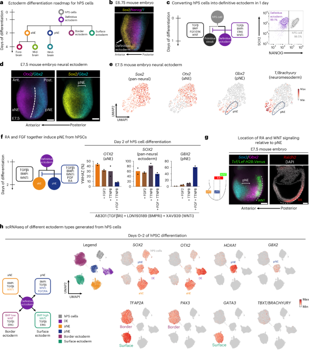
Researchers found that two distinct neural ectoderm progenitor populations — neural plate ectoderm and non-neural ectoderm — each contribute cells to the developing brain. These are not overlapping lineages or transient intermediates. They are parallel origins that coexist during gastrulation and both seed neural structures, including the forebrain and hindbrain.
The finding matters because it exposes an error in the conceptual map scientists have been using to trace cell fate. When your map is wrong, the diseases you search for appear in the wrong places.
What the data actually shows
The study combined single-cell RNA sequencing, bulk transcriptomics, and OmniATAC-sequencing across human pluripotent stem cell-derived models and embryonic tissues. The datasets, deposited under SuperSeries accession GSE286214 at the NCBI Gene Expression Omnibus, reveal molecular signatures that cleanly separate the two progenitor populations across multiple developmental stages.
Gene expression profiles showed that non-neural ectoderm contributes progenitors expressing anterior neural markers, including Otx2, which marks forebrain and midbrain identity. Neural plate ectoderm contributed hindbrain and spinal cord progenitors marked by Gbx2 and Hox genes.
Chromatin accessibility data confirmed these are not transient states but stable, lineage-committed populations with distinct regulatory landscapes. The two progenitor types diverged early in gastrulation and maintained separate epigenetic identities through neurulation.
This is not a borderline finding. The separation is clean across multiple experimental replicates and independent analytical pipelines. Computational scripts used in the analysis are available on GitHub under the Loh laboratory repository.
Why this upends established dogma
The traditional model, established through classical fate-mapping experiments in amphibians, birds, and mammals, treats the neural plate as a single continuous structure. Ectoderm either becomes neural plate or remains non-neural. The boundary between them is sharp. There is no mixing.
That model rests on experiments where labeled cells from specific ectodermal regions were tracked as embryos developed. Those experiments consistently showed neural plate-derived cells populate the neural tube and non-neural ectoderm forms skin and sensory placodes. No study detected significant contribution from non-neural ectoderm to brain tissue.
The new paper does not argue those earlier experiments were flawed. It argues they missed something. The techniques available then — radioactive labels, vital dyes, limited resolution — could not resolve the subtle, early contribution of non-neural ectoderm progenitors to neural structures. Single-cell omics can.
More importantly, the old model treated the neural-non-neural border as a hard boundary. This data shows it is more of a gradient with functional overlap. Cells at the border retain dual potential and can contribute to either lineage depending on signaling context. That changes how we think about competence and fate restriction during early development.
Who wins and who loses
The winners are researchers studying human neurodevelopment. For decades, stem cell protocols for generating brain organoids have assumed a single progenitor origin. Those protocols now need revision. Organoid models that fail to incorporate non-neural ectoderm contributions may be producing incomplete or inaccurate representations of early brain development.
The broader regenerative medicine field also benefits. Understanding that two parallel lineages contribute to brain formation opens new pathways for directing stem cells toward specific neural fates. Protocols that target only the neural plate pathway may be missing a substantial source of progenitor diversity.
The losers are simpler. Textbook authors will need to update chapters on neural induction and early brain patterning. Graduate programs built around the single-origin model will face curriculum adjustments. Any research framework that assumes neural tube derivatives come exclusively from neural plate ectoderm will need reexamination.
What this means for neurodevelopmental disorders
Most neurodevelopmental disorders — autism spectrum disorder, intellectual disability, epilepsy — trace their origins to early brain development. If the brain arises from two progenitor populations rather than one, then mutations affecting neurodevelopment may disrupt either lineage independently or both simultaneously.
This has direct implications for disease modeling. Genetic variants identified in patients with neurodevelopmental conditions may be misclassified if researchers assume all neural cells derive from a single progenitor pool. A mutation affecting non-neural ectoderm-derived brain progenitors would not show up in assays designed only for neural plate-derived cells.
The study’s data provides molecular signatures for both progenitor populations, which means researchers can now design targeted screens for disease-associated variants affecting each lineage separately. This is not speculation. The scRNAseq data is publicly available and can be mined immediately.
The paper also raises questions about conditions like holoprosencephaly and neural tube defects that result from early patterning failures. If two progenitor populations must coordinate their contributions, disruptions to inter-progenitor signaling could produce phenotypes that do not fit existing分类 schemes.
What happens next
The immediate priority is validation. The findings need confirmation in human embryonic tissues at comparable developmental stages, not just stem cell derivatives. Fate-mapping studies using modern genetic tools should be able to test this directly in model organisms.
The second priority is integrating the two-origin model into existing frameworks of brain regionalization. The midbrain-hindbrain boundary, already studied extensively through work on Otx2 and Gbx2 interactions, takes on new meaning if both progenitor populations contribute to its formation.
Organoid researchers need to revise protocols. The field has invested heavily in models based on the single-origin assumption. Changing course will cost time and resources but avoiding the correction will waste far more.
Perhaps most importantly, this finding demonstrates that even well-established developmental paradigms can contain blind spots large enough to reshape entire fields. The neural plate has been studied since the nineteenth century. New technology revealed what decades of careful observation missed. The same may be true for other areas of developmental biology where accepted models rest on older experimental limitations.
The data is public. The mechanisms are now testable. The old textbook story is over.