The brain is not two brains, but it is two territories
A Stanford-led study shows the hindbrain and spinal cord inherit their identity from cells that know their place before neural tissue even forms. The takeaway is less about how many brains you have and more about how developmental biology sets the rules for research.
The headline was wrong. The science is not.
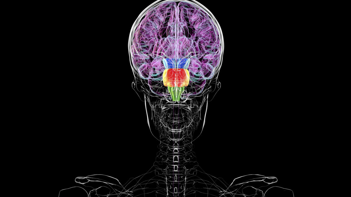
You do not have two brains. The phrase made rounds across popular-science outlets last week after a press release from Stanford described a new study suggesting the hindbrain and spinal cord carry an early developmental fate separate from the midbrain and forebrain. A single fluorescent tag made the boundary vivid: red in the hindbrain, cyan everywhere else. A sound bite is not a finding.
But the underlying work, led by researchers at Stanford and published in Nature, is substantive. It traces how vertebrate embryos split a sheet of ectoderm into distinct neural territories long before neurons fire or any recognizable brain structure appears. The finding matters because it reframes how neuroscience thinks about regional identity, how medicine might direct stem cells toward specific brain regions, and how computational models of neural development can move beyond treating the brain as a homogeneous substrate.
What the study actually shows
The team engineered mice so that early ectoderm cells expressed fluorescent proteins wherever specific regulatory genes were active. Half the embryonic ectoderm glowed red, half glowed cyan. As the neural tube formed, that color map persisted. The hindbrain and spinal cord stayed red. The rest of the brain stayed cyan. In 96 percent of single-cell labeling experiments, descendants of one tagged cell ended up entirely in either the hindbrain or the mid- and forebrain — never split across both.
Human stem cells showed the same pattern when exposed to the same early signals. Nerve cells inherit positional information from the tissue they form from. If you want hindbrain neurons, you have to send stem cells down the hindbrain path first. The boundary is established before the neural tube even fully closes.
That 96 percent figure is worth noting. It is not 100 percent, which means either the experimental system has limits or cells near the ectodermal border retain enough plasticity to change fate if they shift position during a narrow developmental window. The authors do not resolve that ambiguity. Equally underexplored is the boundary between midbrain and hindbrain at the cellular level — the paper cites a single prior reference suggesting early segregation, and calls for more work.
Why early segmentation matters
The nervous system develops as a simple tube. Early on, almost every cell in that tube is exposed to the same two signaling molecules: sonic hedgehog from the ventral side and BMPs from the dorsal side. If every neural precursor responded identically, the brain would be a far simpler organ.
Segment identity solves that problem. Cells with different developmental histories interpret the same signals differently. Midbrain cells respond to BMPs in ways that spinal cord cells do not. The same signal gets repurposed across developmental stages — first to specify whether a cell becomes neural at all, then to assign regional identity, then to guide neuron migration. Evolution does not need entirely new signaling systems for each new function. It reuses existing ones and lets context determine the output.
Boundaries between segments are not passive lines. At the midbrain-hindbrain boundary, cells activate a distinct set of genes that produce signaling molecules affecting tissue on both sides. Those signals trigger different responses depending on the developmental history of the neighboring segment. Boundaries are generative, not just demarcating.
Who wins from this reframe
Neuroscientists studying regional specialization gain a clearer model. Instead of asking how a fully formed brain divides its functions, they can ask how early positional information constrains later differentiation. That shifts the question from architecture to lineage.
Regenerative medicine benefits directly. Stem cell protocols for generating specific neuron types — dopaminergic cells for Parkinson’s, motor neurons for ALS — depend on recapitulating developmental signals in the right order. This work provides evidence that early segment identity is non-negotiable. You cannot skip from a generic neural precursor to a hindbrain neuron without passing through the right early signals. Protocols that ignore that sequence will produce the wrong cells, or none at all.
Aging research gains a methodological lens. If segment identity is established early and maintained, then age-related decline may involve degradation of those early programs, not just accumulation of damage. That opens a different set of therapeutic questions than the standard damage-repair frame.
Artificial intelligence researchers should pay attention, too. Most neural network architectures treat layers as functionally interchangeable blocks. A developmentally informed approach would explore whether early architectural segregation — distinct modules with different initial conditions — produces systems that learn more efficiently and generalize better. The brain does not solve the modularity problem by starting uniform and dividing later. It divides first and builds from there. That ordering may matter for AI as much as for biology.
What is still unknown
The study maps one boundary with high resolution. The mechanisms that establish the midbrain-forebrain border remain poorly understood. The relationship between hindbrain and spinal cord fates has only one prior reference to build on. How the 4 percent of cells that cross the labeled boundary behave — and whether they are noise or biologically meaningful — is unresolved.
The timing is also unclear. The fluorescent tags report gene activity at a snapshot, but development happens continuously. The authors note the difficulty of correlating what they see with the actual timing of gene expression changes. That is a technical limitation, not a conceptual one, but it leaves room for revision as live-imaging methods improve.
The real story
The popular framing — two brains — collapses a nuanced developmental finding into a click line. The actual story is more interesting. A single sheet of cells knows where it is along the head-to-tail axis before it ever becomes neural tissue. That positional information is inherited, maintained, and amplified as the nervous system grows. Early segmentation is not a curiosity. It is the mechanism that allows a limited toolkit of signals to generate a complex brain.
The take-away for researchers is not that the brain is two organs. It is that region identity is established before function exists, and that identity shapes everything that follows. Treat the brain as a unified mass and you miss the constraints that make it work. Treat it as a collection of early territories and you start asking the right questions.