Why the Stanford Human-Mouse Brain Study Isn't Just Another Xenograft Milestone
Stanford's new model — human brain tissue growing inside mice missing most of their cortex — looks like a neuroscience breakthrough. The real story is how it resurrects the same ethical fault lines that have shaped biomedical funding for decades.
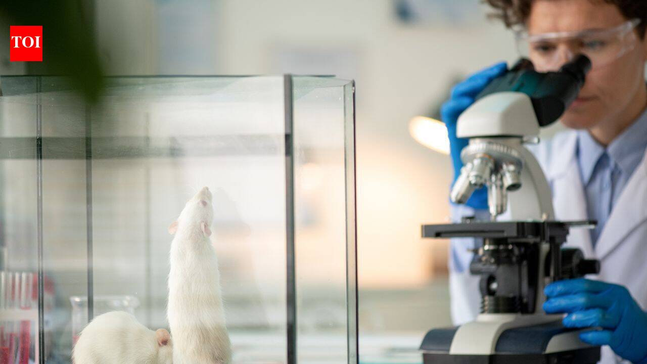
The imagery does the work for you
Human brain tissue growing inside a mouse. Not floating in a petri dish — integrated. Synapses firing. Taking up over 90 percent of the cortical cavity after three months. The cover-image potential of this study is almost too on-the-nose: a humanized mouse brain, a living hybrid.
That is why the publication of “Developmental xenocortication using human-derived organoids in mice” in Nature on September 16 is already generating noise beyond the neuroscience circles that normally chase these papers. It is not the first time human cells have been grafted into rodents. It is not even the first time cortical organoids have been transplanted into animal hosts. Stanford’s apallial mouse — engineered to lack most of its neocortex from the start — simply removed the competition that had limited previous attempts.
What makes this study ethically combustible is the scale of the takeover, not the novelty of the technique.
How the model works
Sergiu Pasca’s team at Stanford Medicine has spent more than a decade refining cortical organoids — three-dimensional clusters of human brain cells grown from reprogrammed skin stem cells. Organoids self-organize into structures that mimic developing cortex, but they are starved of blood supply, immune input, and long-range connections when kept in a dish.
Earlier work transplanted these organoids into newborn rats. The human neurons grew larger, branched more extensively, and fired more electrically. But the rat’s own cortex developed fast and crowded the graft out. The human tissue never got enough room to fully integrate.
The apallial mouse solves that problem. These mice carry a genetic edit that prevents starter cells for most of the neocortex from forming. Adults have roughly 2 percent of the cortical tissue found in a normal mouse. That leaves a large empty space — a biological niche waiting to be filled.
When two-month-old human cortical organoids were transplanted into two-day-old apallial pups, the human tissue survived, expanded, and took over. By three months, more than 90 percent of the cortical volume was human. The neurons projected axons, formed synapses, and connected to the mouse’s remaining brain regions and spinal cord.
Behavioral testing at three to six months showed the xenocortical mice moving and behaving broadly like normal mice, with subtle gait and memory differences that suggest the full cortex has a stabilizing role.
Why this matters for disease research
The model’s power comes from its specificity. Because the organoids carry the donor’s genetic material, researchers can now grow human cortex from patients with autism, epilepsy, or schizophrenia and watch those disease-associated circuit changes unfold in a living, behaving animal.
In one proof-of-concept experiment, the xenocortical mice were exposed to five hours of low oxygen. The human cortical tissue suffered substantial damage, and the mice showed balance and movement problems resembling cerebral palsy. Normal mice and apallial mice without human tissue were largely unaffected. The difference points to a vulnerability unique to human cortical neurons under oxygen stress — a clue that could help explain why premature babies and children born with birth asphyxia are disproportionately affected.
Perhaps the most striking finding was the emergence of von Economo neurons — large, cigar-shaped cells linked to social awareness and decision-making — inside the transplanted human tissue. Until now, these cells had only been seen in post-mortem human brains or in other large-brained social animals. They had never appeared in lab-grown organoids or earlier rodent transplant models. Their spontaneous appearance in the xenocortical mice suggests the living, integrated environment may be necessary for certain rare human neuron types to mature at all.
That has direct implications for frontotemporal dementia and other disorders where von Economo neurons degenerate early.
The ethical fault line, reopened
Work that blends human brain tissue with animal brains has always raised questions. The Stanford team has consulted ethicists, neurobiologists, patient advocates, philosophers, and legal scholars for years. In late 2025, Pasca convened a conference at Asilomar — the same California retreat where AI safety researchers now gather — to debate the implications of using human stem cell models in this way.
The central argument in favor is straightforward: hundreds of millions of people live with untreatable neurological disorders, and current models fail to capture the human-specific biology that drives these diseases. Animal models of mouse or rat cortex do not translate neatly. Organoids in a dish lack the systemic context that shapes how neurons actually develop. If a hybrid model can reveal mechanisms that neither system shows alone, the moral case for using it is strong — especially when the animals involved are engineered to have minimal native cortex and show no evidence of altered consciousness.
But the imagery is the problem. A mouse brain that is 90 percent human cortex looks, even to a scientifically literate audience, like something out of a thriller. The Asilomar conference itself signals that the researchers feel the weight of that perception. They are not dismissing concern; they are trying to get ahead of it.
What this means for funding and regulation
Stanford has patents on aspects of the technology. The team expects other labs to build on the approach. Within a few years, xenocortication could become a standard model for studying human-specific neurological conditions — and with that adoption will come renewed scrutiny from ethics review boards, funding agencies, and the public.
The broader pattern is familiar. Every time a technique crosses a visible threshold — whether it is humanized primates, organoid-awareness debates, or now apallial mice with extensive human cortex — the same cycle repeats: scientific advance, ethical alarm, policy response, funding recalibration. The difference this time is the speed. Social media amplifies the imagery instantly. The Asilomar reference, borrowed from the AI safety community, shows how bioethics and AI ethics are increasingly sharing the same vocabulary and the same anxiety.
The question is not whether this research will continue — it already has patents and a Nature paper behind it. The question is what guardrails get built around it, who gets to define them, and whether the regulatory conversation moves fast enough to keep pace with the science.
For now, the xenocortical mouse is a tool for studying disease. It is not a model of human consciousness. But the fact that it looks the way it does means the ethical debate will not stay inside academic journals.